
Inside the Bite: The Komodo Dragon
Inside the Bite: The Komodo Dragon
Varanus komodoensis is the world’s largest living lizard. Known as the Komodo dragon because of its enormous size, a fully grown Varanus komodoensis can exceed 3 meters in length and 70 kilograms in weight. It can only be found on a few islands in Indonesia, and is considered vulnerable by the IUCN because it has been eliminated from most of its range by humans. As of today, only about 4,000 of these magnificent reptiles remain in the wild (1). This is unfortunate because Western science only “discovered” the species in 1910 (2). Despite its conservation status the Komodo dragon dominates the islands of Komodo, Flores, Rinca, Gili Dasami, and Gili Montang, as the apex predator of its ecosystem.

RANGE OF THE KOMODO DRAGON IN INDONESIA (2)
Although primarily a scavenger, the Komodo dragon also hunts invertebrates, birds, and mammals. It can also take down humans, deer, and water buffalo (a large invasive species introduced by humans) many times its size with a single bite. The mechanisms that make this feat possible have only been discovered by scientists over the past fifteen years. The key

is highly toxic saliva. In 1996 and 1997, a group led by Texas businessman Terry Fredeking and biologist Joel Montgomery took blood and saliva samples from 23 wild Komodo dragons and similar samples from 13 captive dragons. Since then, they’ve analyzed the saliva samples and found at least 57 species of bacteria (3). Below is a table taken from that study, displaying the species of bacteria extracted from those dragon samples. Prominent pathogens found include Escherichia coli, Pasteurella multocida, Enterobacter cloacae, Proteus morgani, and P. mirabilis. Numerous Staphylococcus, Streptococcus, and Bacillus species were present as well. Along with illustrating the pathogenic diversity of the Komodo dragon mouth, the study also bolstered the observation that wounds from dragon bites cause septic shock and bacterial infections in prey.
Conditions of the Komodo Dragon’s Mouth
Life inside a dynamic environment is never easy for the organisms that call it home. The Komodo dragon’s mouth is not unique to the reptile world; most reptiles have mouths with similar internal conditions. Its pH is close to neutral, while its temperature varies over the course of the day because the Komodo dragon is ectothermic. However, despite this ectothermy it has a relatively stable core body temperature (5). On top of that, these dragons are born with clean and sterile mouths and are responsible for developing their own bacterial flora (6). As they devour carrion, they collect bacterial colonies from decaying animal flesh. Pieces of meat get caught in between their 60 serrated teeth, feeding the bacteria a reliable source of nutrients. In addition, the teeth provide thermally stable surfaces that remain at a constant, optimal 37 degrees Celsius (7). Komodo dragons also expose themselves to other bacteria in the water, soil, and feces, creating a caustic cocktail that can cripple their prey.
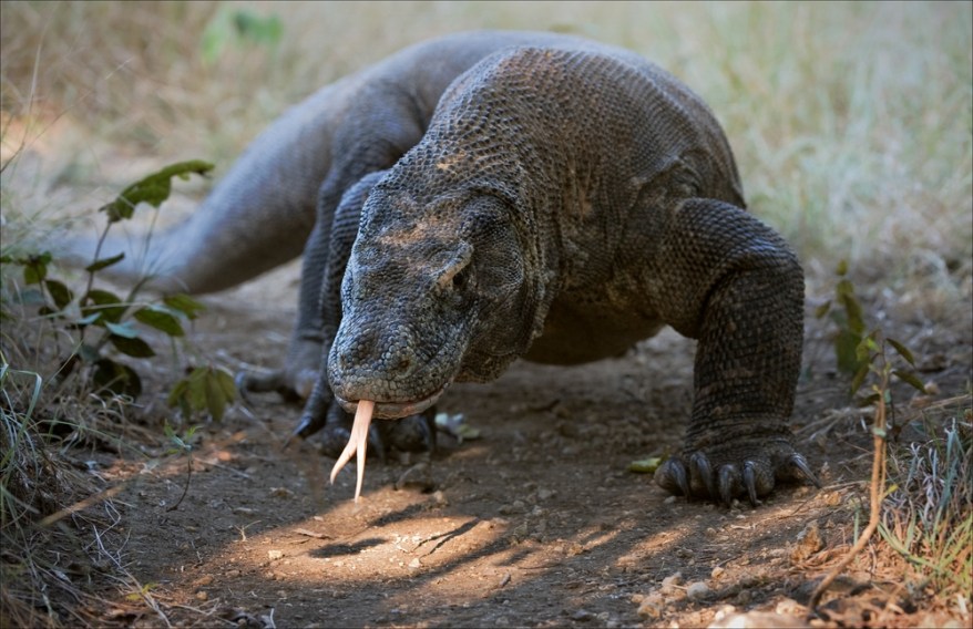
Komodo dragons have long, forked tongues that they use to help smell and taste.
Credit: Sergey Uryadnikov / Shutterstock
E. coli in the Dragon’s Mouth
Escherischia coli is, perhaps, the best-described bacterium in the Komodo dragon’s arsenal. Its genome has been completely sequenced many times, and is the model bacterium for Western science. E. coli usually inhabits the lower intestine of warm-blooded organisms, but strains of this species have been found elsewhere in a wide variety of environments. Most strains are harmless, essential for the proper digestion of food. Using pili, the bacterium adheres to the mucus that lines the walls of the large intestine. A facultative anaerobe, E. coli can function properly with or without oxygen. It is the primary facultative anaerobe of the human gastrointestinal tract, and is important to the Komodo dragon as well. E. coli is Gram-negative, facultative anaerobic and non-sporulating. Cells are typically rod-shaped and are about 2 micrometres (μm) long and 0.5 μm in diameter, with a cell volume of 0.6 – 0.7 μm3 (8). They can also survive on a wide variety of substrates, breaking down nutrients via mixed-acid fermentation when oxygen is absent. This produces lactate, succinate, ethanol, acetate, carbon dioxide, and sometimes hydrogen gas. E. coli grows best at 37 degrees Celsius (9), though it can survive for prolonged periods at a wide variety of temperatures. E. coli possesses a Gram-negative cell wall. This means 10% of its cell wall consists of peptidoglycan, the structural polysaccharide that gives structure to most prokaryotic cells. It also means that the bacterium contains two bilayers, an inner one and an outer one that can be very toxic to predators. Virulent strains of E. coli take advantage of this property, inflicting gastroenteritis, urinary tract infections, and neonatal meningitis upon immunocompromised prey animals.
The most virulent species found in Komodo dragon saliva are Pasteurella multocida and Pseudomonas aeruginosa. These two species work together to create the dragon’s potent and infamous bite. Amongst the 57 identified bacteria found in the mouth of the Komodo dragon, Pasteurella multocida was determined to be amongst, if not the main culprit in bringing down any prey (10). Its co-conspirator is Pseudomonas aeruginosa, which will be discussed later.
Key Infective Agents in Dragon Saliva: Pasteurella multocida
Pasteurella multocida resembles E. coli, being both Gram negative and a facultative anaerobe. It is typically found in the respiratory tracts of livestock, poultry, and domesticated pet species (11) in a symbiotic relationship, when not found in the mouth of a Komodo dragon. It grows best at 37 degrees Celsius. It has not been as thoroughly studied as E. coli; scientists have yet to sequence its genome. Being a facultative anaerobe, it is oxidase-positive and catalase-positive, and can also ferment a large number of carbohydrates under anaerobic conditions (12).
A bacteriophage causes virulence in P. multocida. The virus encodes the toxin responsible for most P. multocida virulence factors. This toxin activates Rho GTPases, which bind and hydrolyze GTP, and are important in actin stress fiber formation. Formation of stress fibers may aid in the endocytosis of P. multocida. The host cell cycle is also modulated by the toxin, which can act as an intracellular mitogen (13). This stress fiber formation causes swelling on the epidermis, which allows the bacteria to enter the host’s cells (11). This means that even if the prey initially escapes the Komodo dragon after it has been bitten, it will fall to serious infection in a few days. If the infection is restricted to the area of infection on top of the epidermis typically from a bite, rapid progression of cellulitis or rash formation, swelling, redness, tenderness, and pain in the area, followed by fevers, chills, and headaches are observed. Though typically not fatal, P. Multocida can have more detrimental effects if infecting elsewhere (14). P. Multocida can also cause respiratory problems where the upper respiratory tract can become inflamed, making breathing difficult and painful. In more serious locations, cardiovascular problems can arise where the infected host suffers from inflammation of the heart tissue and disturbance in normal heart rhythm—both of which can be lethal. To make matters worse, if the bacteria are able to enter the central nervous system and cross the blood-brain barrier, they can trigger meningitis, or inflammation of the protective tissue of the brain (11). This inflammation would cause many of the prey’s vital organ systems to shut down after a certain length of time.
Key Infective Agents in Dragon Saliva: Pseudomonas aeruginosa
Pseudomonas aeruginosa is also Gram negative, but relies on oxygen to survive. Although classified as an aerobic organism, P. aeruginosa is considered by many as a facultative anaerobe, as it is well adapted to proliferate in conditions of partial or total oxygen depletion. This organism can achieve anaerobic growth with nitrate as a terminal electron acceptor, and, in its absence, it is also able to ferment arginine by substrate-level phosphorylation (15). Numerous strains have had their genomes sequenced because P. aeruginosa can be found anywhere. It has a tolerance for a wide variety of environmental conditions both inside and outside a host. This simple Gram negative, aerobic rod shaped bacterium, measuring 0.5 to 0.8 micrometers to 1.5 to 3.0 micrometers, has rather simple nutritional requirements—it can grow even if the medium is distilled water. Moreover, its metabolism is extremely versatile, as it does not necessarily need growth factors and can use more than 75 organic compounds for biosynthesis (16). It can use nitrate as a primary electron acceptor during cellular respiration when oxygen is not present, allowing this aerobic organism to thrive under anaerobic conditions.
As a free-living organism, P. aeruginosa can thrive anywhere. As a parasite, like after a Komodo dragon bite, the bacterium is indestructible. Once a Komodo dragon crunches off a nice chunk of flesh off the prey, aside from inducing massive blood loss, the Komodo compromises the skin—which serves as the pivotal external barrier against bacteria—and allows the bacteria safe passage into the prey. Once tissue defenses are compromised, there’s no tissue—spare a few—P. aeruginosa can’t infect; it can cause infections in the urinary tract, respiratory system, soft tissue, bone and joints, and gastrointestinal region to name a few, and trigger bacteremia and septicemia (septic shock).
Once inside an infected host, P. aeruginosa can disassociate from the biofilms on the teeth and invade healthy tissues. Their incredible growth can be attributed to several factors. For one, it is one of the most vigorous, fast swimming bacteria via a single polar flagellum. Secondly, it encodes a mucoid capsule—possibly lipopolysaccharides—to avoid phagocytosis and related bacterial responses. Thirdly, the bacteria produce two extracellular proteases in the form of elastase and alkaline protease; the elastase effectively cleaves host antibodies, collagen, and related complement and lyses fibronectin, exposing receptors for attachment on the mucosa of tissues; the alkaline protease aids elastase in lysing fibrin and disrupting the formation of fibrin. Fourthly, P. aeruginosa produces three soluble proteins in the form of a pore-forming cytotoxin and two hemolysins in the form of phospholipase and lecithinase—both work synergistically to break down lipids and lecithin. Its virulence doesn’t end there. Fifthly, it releases two extracellular protein toxins—exoenzyme S and exotoxin A. Exoenzyme S functions to impair phagocytic cell activity in the bloodstream and internal organs to set the stage for invasion. Exotoxin A inhibits protein synthesis in its target cell by causing ADP ribosylation of eukaryotic elongation factor 2 (14).
The Biofilm Inside the Komodo Dragon’s Mouth
The species mentioned above provide only a snapshot of the vast microbial diversity of the Komodo dragon’s mouth. The 50 to 80 species that call the lizard’s oral cavity home form multi-specie colonies on the surface of its teeth via fimbrial adherence (pili) as a means of maintaining homeostasis and resisting phagocytic and antimicrobial elements, pH changes, and fluctuations in temperature. Their resilience to external pressures and conditions is due to an exopolysaccharide coat, which is synthesized by glycosyltransferase enzymes on the bacterial cell’s exterior. This coat is porous and has appropriate channels for nutrient uptake (17) (4). The cells communicate with each other via quorum sensing (4).
By comparison, the human oral cavity houses hundreds of species of bacteria, viruses, and fungi. Many of these can associate to form biofilms, which are resistant to mechanical stress or antibiotic treatment. Most are also commensal species, but they can become pathogenic in responses to changes in the environment or other triggers in the oral cavity, including the quality of an individual’s personal hygiene (20). The Komodo dragon, however, seems unaffected by the pathogens in its body.
Conclusion
Recent surprises in research suggest that microbes are not entirely responsible for Varanus komodoensis’s extraordinary bite. In 2009, scientists at the University of Melbourne examined preserved dragon skulls and discovered complex venom glands and specialized serrated teeth which create deep lacerations for entry of the venom. The effects of venom were also tested by the team and found to be similar to that of the gila monster and many snakes which cause a severe loss in blood pressure by widening blood vessels, thereby inducing shock in a victim. These findings may explain the observations by Dr. Fry and others that Komodo dragon prey become still and unusually quiet soon after being bitten. Bitten prey also bleed profusely, consistent with the team’s discovery that the venom was also rich in toxins that prolong bleeding (18). The Komodo dragon surprised scientists again in 2006 when a female specimen in a French zoo underwent a “virgin birth”. Through parthenogenesis she fertilized her own eggs, producing viable male offspring. In effect, she acted as both a mother and father to her offspring (19).
The Komodo Dragon is a remarkable beast that has terrorized man’s imagination since its discovery. A recent addition to Western science, it seems that the Komodo dragon is only recently making headlines. Amazing discoveries involving this amazing creature have been made over the past decade, and more will surely follow as long as it is protected from extinction.
Works Cited
- Lopata, Peg. “The Drooling Dragons of Komodo Island”. Faces (07491387); May/Jun2009, Vol. 25 Issue 8, p6-7
- Ciofo, Claudio. “The Komodo Dragon”. Scientific American. March 1999. pp. 84-91
- Feldman, Ruth Tenzer. “Dragon Drool!”. Odyssey; Feb2007, Vol. 16 Issue 2, p. 49
- Montgomery, Joel M., et. al. “Aerobic Salivary Bacteria in Wild and Captive Komodo Dragons”. Journal of Wildlife Diseases, 38(3), 2002, pp. 545–551
- Lutz, Dick and J. Marie Lutz. Komodo, the Living Dragon: Living Dragon. Salem: DIMI Press, 1997
- “Explore by Animal Faust: Into the Dragon’s Mouth.” Animal Stories. 2008. Shedd The World’s Aquarium. 29 Aug 2008
- Young, Emma. “Lizards’ Poisonous Secret is Revealed.” NewScientist. Nov. 2005.http://www.newscientist.com/article.ns?id=dn8331
- Kubitschek HE (1 January 1990). “Cell volume increase in Escherichia coli after shifts to richer media”. Bacteriol. 172 (1): 94–101
- Fotadar U, Zaveloff P, Terracio L (2005). “Growth of Escherichia coli at elevated temperatures”. J. Basic Microbiol. 45 (5): 403–4.
- Rogers A H. (2008). Molecular Oral Microbiology. Caister Academic Press
- Cohen, Jesse. Smithsonian National Zoological Park. http://www.amnh.org/exhibitions/lizards/images/komodo_smithsonian.jpg
- Casolari C, Fabio U. “Isolation of Pasteurella multocida from Human Clinical Specimens: First Report in Italy”. European Journal of Epidemiology. Sept 1988; 4(3):389-90
- Lacerda HM, Lax AJ, Rozenqurt E. “Pasteurella multocida toxin, a potent intracellularly acting mitogen, induces p125FAK and paxillin tyrosine phosphorylation, actin stress fiber formation, and focal contact assembly in Swiss 3T3 cells”. J Biol Chem. 5 Jan 1996; 271(1):439-45.
- “Komodo Dragon Mouth Niche”. MicrobeWiki. 20 August 2010. 19 November 2010. http://microbewiki.kenyon.edu/index.php/Komodo_Dragon_Mouth_Niche#Location_and_Physical_Conditions
- Collins FM (1955). “Effect of aeration on the formation of nitrate-reducing enzymes by aeruginosa“. Nature 175 (4447): 173–4
- “Komodo Dragon”. org. 20 November 2010. http://en.wikipedia.org/wiki/Komodo_Dragon
- Lafeber, Thomas, MD., J. Robert Cantey, MD. “Pasteurella Multocida Infections.” eMedicine. April 2006. http://www.emedicine.com/MED/topic1764.htm
- “University of Melbourne; A combined tooth-venom arsenal revealed as key to Komodo dragon’s hunting strategy”. Telemedicine Business Week. Atlanta: Jun 3, 2009. 274
- “Zoo awaits dragon’s ‘virgin’ birth: In an evolutionary twist, Flora the Komodo dragon has managed to become pregnant all on her own, without any male help”. Ottawa Citizen. December 21, 2006. NEWS; Pg. A6
- Maria Avila, David M. Ojcius, Özlem Yilmaz. DNA and Cell Biology. August 2009, 28(8): 405-411. doi:10.1089/dna.2009.0874.
WordPress Community Snapshot
Recent Comments
| Anonymous on On the Sharks of the Devo… | |
| Mariam Weber on Off Topic: How Rent Works in… | |
| Marius on The Shadow Paintings | |
| Arp ad on Community Conversations: Intro… | |
| Autism and The Limit… on The Autistic Voice |

Recent Comments